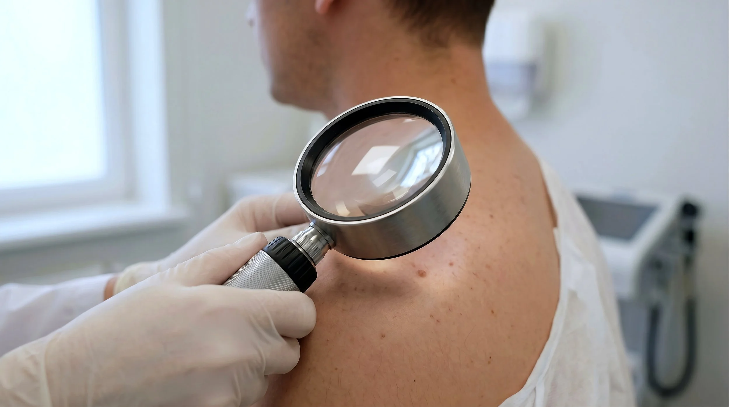
skin cancer & Dermoscopy
— THE TECHNOLOGY
What Is a Dermoscopy?
Dermoscopy uses polarised light and magnification to examine the deeper layers of skin - structures invisible to the naked eye. In trained hands, it significantly improves diagnostic accuracy, helping catch early-stage melanomas that would otherwise go undetected.
Dr Ahmed holds a Professional Certificate of Dermoscopy and uses it in every skin cancer consultation. No referral needed - available at all three clinics.
-
Systematic examination of all skin surfaces, flagging any lesions for closer review.
-
Polarised imaging reveals subsurface structures invisible to the naked eye.
-
Monitor, biopsy, or excise explained clearly before you leave.
-
Where needed, excision is often performed the same day under local anaesthetic.
The Three Main Skin Cancers
-
![]()
Basal Cell Carcinoma
MOST COMMON · SLOW GROWING · RARELY SPREADS
Grows slowly and rarely spreads, but causes local damage if untreated. Often looks like a pearly bump or flat scar-like patch - not what most people expect skin cancer to look like. -
![]()
Squamous Cell Carcinoma
SECOND MOST COMMON · CAN SPREAD · TREAT PROMPTLY
Arises on sun-damaged skin and carries a higher risk of spreading than BCC. Often appears as a firm red nodule or a scaly, crusted lesion that won't heal. -
![]()
Melanoma
MOST DANGEROUS · CAN BE AGGRESSIVE · EARLY DETECTION CRITICAL
Less common but far more serious. Can spread rapidly if not caught early. When detected at stage one, survival rates exceed 95% — which is why regular checks matter.



— WHAT I OFFER
Skin Cancer Services
-
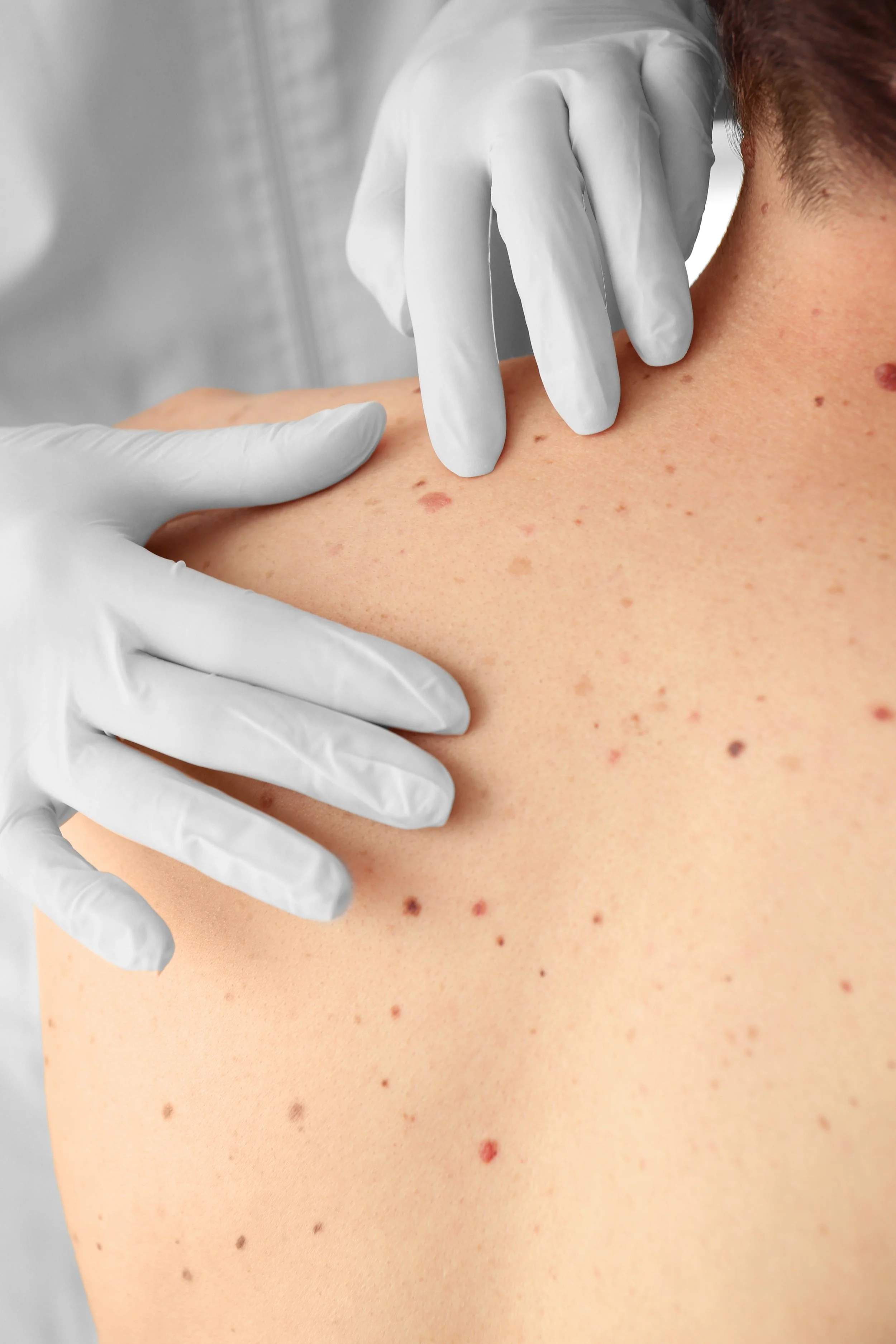
Systematic head-to-toe examination with dermoscopic imaging of any suspicious lesions.
-
Targeted polarised imaging for lesions that need a closer look - no referral needed.
-
Certified in-clinic surgical removal of BCCs, SCCs, and melanomas including face and scalp.
-
Punch or excisional biopsy sent to pathology for definitive diagnosis.
-
Liquid nitrogen treatment for actinic keratoses and pre-cancerous skin lesions.
-
Structured monitoring every 6–12 months for patients with elevated skin cancer risk.
— THE TECHNOLOGY
Who Should Get Checked?
In Australia, the question isn't whether you're at risk; it's how high. If any of these apply to you, don't put it off.
How Often Should You Get Checked?
Most adults: Once a year as a baseline.
Elevated risk:Every 6–12 months depending on your history.
Prior melanoma: Every 3–6 months with dermoscopic monitoring.
New or changing lesion:Don't wait - book a targeted consultation promptly.